Service
Breast surgery
A lump you can feel, pain, discharge or a finding picked up on imaging may need surgical assessment. The aim is not to operate on every finding — first to establish what we are dealing with, then to decide whether an intervention is needed at all.
On this page
Conditions covered
Most breast pathology is benign. The list below covers the diagnoses seen most often in clinic — the assessment pathway and the indication for surgery differ for each of them.
- Benign breast disease: fibroadenoma, phyllodes tumour, intraductal papilloma, cyst, breast abscess, accessory breast tissue
- Benign breast tumours and benign nodules that recur, grow or cause discomfort
- Malignant nodules and breast cancer — surgical treatment at every stage
- Inflammatory conditions: mastitis and idiopathic granulomatous mastitis
- Soft tissue tumours — lipoma and similar lesions in the breast area and elsewhere
Who comes to the clinic
- People who have felt a lump or an area of firmness in the breast
- Women with a suspicious finding on ultrasound, mammography or MRI
- Anyone with bloody or clear discharge from the nipple
- Skin dimpling, puckering or an “orange peel” appearance
- Inflammatory conditions with persistent redness, swelling and pain
- People who have a biopsy result and need a surgical decision
- Those with breast or ovarian cancer in the family who want a risk assessment
Breast disease also occurs in men — rarely, but any lump found in a man must be investigated.
How the assessment works
-
Conversation and physical examination
When the symptom started, whether it changes with the menstrual cycle, family history and current medication are established. The breasts and the axillae are then examined.
-
Imaging
Depending on age, ultrasound, mammography or both are used. Elastography or MRI may add information in some cases. Imaging alone does not make a diagnosis — the finding is read together with the clinical picture.
-
Biopsy, when needed
For suspicious findings a tissue sample is taken (fine needle aspiration or core needle biopsy). The pathology result is the basis of the treatment plan. A biopsy does not mean “there is cancer” — it is very often done precisely to show that there is not.
-
Making the decision
Many benign findings are simply monitored. Whether surgery is needed depends on the type of lesion, its size, how fast it grows, the symptoms and the pathology result.
Which operations are performed
- Removal of benign lesions — fibroadenoma, phyllodes tumour and similar lesions when they grow, change quickly or cause discomfort
- Treatment of cysts — needle aspiration of simple cysts, surgical removal for recurrent or complex ones
- Removal of intraductal papilloma — surgical exploration and excision of the duct (microdochectomy) where there is bloody nipple discharge
- Treatment of a breast abscess — drainage, assessment of the inflammatory focus and a surgical solution for recurrent cases
- Removal of accessory breast tissue — additional breast tissue in the axilla causing pain, swelling or cosmetic concern
- Surgical treatment of breast cancer — breast-conserving surgery (lumpectomy) or mastectomy, assessed individually in every case
- Assessment of the axillary lymph nodes — sentinel lymph node biopsy (SLNB) or axillary lymph node dissection (ALND)
- Removal of soft tissue lesions — lipoma and similar lesions in the breast area and on the chest wall
A research interest
Inflammatory breast conditions, in particular idiopathic granulomatous mastitis, are among the surgeon's research subjects and feature in his work published in international journals.
The oncoplastic approach
Oncoplastic surgery aims to preserve the shape of the breast as far as possible while keeping oncological safety intact. Reshaping the remaining tissue after the tumour has been removed is the core of the approach.
Whether it suits a particular patient depends on the size of the tumour, its position in the breast, the volume of the breast and the additional treatments planned. Suitability is assessed individually at the consultation.
Training in this field includes the 2nd Advanced Oncoplastic Breast Surgical Skills Course at the Nasser Institute for Research and Treatment in Cairo (26–27 April 2025; RCSEd, 12 CPD hours) and the Conservative Mastectomy and Breast Reconstruction Course (ALEXIA) at the Alexandria Faculty of Medicine, Egypt (2026). At the PCCM’S-ISOS & ABDSA oncoplastic and reconstructive breast surgery workshop in Baku (2026, 30 CME credits) the Doctor spoke on incisions and techniques in nipple-sparing and skin-sparing mastectomy. See the documents →
Axillary lymph nodes
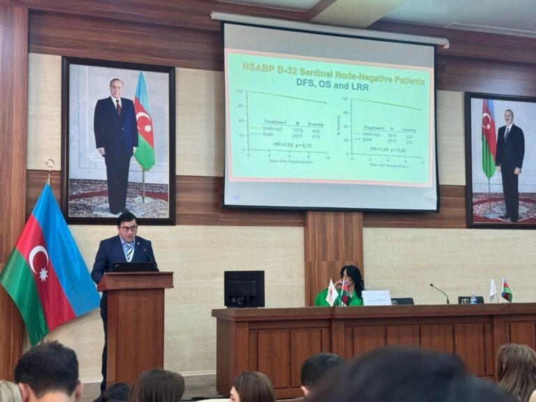
In breast cancer, the state of the axillary lymph nodes directly shapes the treatment plan. Rather than removing all of them, where possible the sentinel lymph node is examined first — the first node the tumour drains into is identified, removed and examined on its own. If it is clear, a wider operation may be avoided.
Axillary surgery after chemotherapy requires separate planning; this was the subject of the surgeon's presentation at the National Congress on Breast Disease in Azerbaijan.
After the operation
The length of stay and recovery vary with the extent of the operation. After smaller procedures it may be possible to go home the same day or the next; wider operations need a few days of observation.
After surgery the pathology report on the removed tissue is awaited. That report determines the next steps — whether further treatment is needed. In cancer cases the plan is drawn up jointly by the surgeon, the oncologist and the radiologist.
Do not delay if you have
- A rapidly enlarging lump or area of firmness in the breast
- Severe redness, swelling and pain with a fever
- Bloody discharge from the nipple
- Broken skin or an ulcer that will not heal
- A newly appeared firm lump in the axilla
For general emergencies, call 103.
Frequently asked questions
I have found a lump in my breast — does that mean cancer?
No. A significant proportion of breast lumps are benign — fibroadenoma, cysts and inflammatory changes among them. But the type of lump cannot be determined by examination alone, so every new finding should be investigated with imaging and, where needed, a biopsy.
Can a biopsy cause cancer to spread?
Current medical evidence does not support this concern. Biopsy is a standard step in diagnosis, and without an accurate diagnosis a correct treatment plan cannot be made. Refusing a biopsy can delay the diagnosis.
Does the whole breast always have to be removed?
No. Breast-conserving surgery is possible in many cases. The decision depends on the size and position of the tumour, the volume of the breast, the number of tumour foci and the additional treatments planned, and is made individually for each patient.
How long before I can go back to work after surgery?
That depends on the extent of the operation and on the nature of your work. Return can be quick after small procedures, while wider operations need longer recovery. The exact timing is discussed individually before the operation.
When should mammography start?
The age at which screening starts depends on your level of risk. Women with breast cancer in the family may be advised to start earlier. A schedule that suits you is agreed at the consultation.
Is granulomatous mastitis breast cancer?
No, it is an inflammatory condition. But because it can mimic cancer on imaging and even on examination, telling the two apart is sometimes difficult and often requires a biopsy.
Updated: